The Science of Ultrasound-Guided PRP: Why Blind Injections Fail

By Dr. Benjamin DuBois
If you are an athlete or an active adult living in San Diego, your shoulders are central to everything you do. Whether you are paddling out at Tourmaline, serving an ace on the tennis courts in La Jolla, or swinging for the fairway in Torrey Pines, you rely on the incredible mobility of your shoulder joints.
When chronic shoulder pain sets in, it does not just ruin your athletic performance—it alters your entire quality of life. Simple, everyday actions like reaching for a seatbelt, lifting a grocery bag, or simply trying to find a comfortable sleeping position at night become exercises in frustration.
When rest, physical therapy, and temporary modifications fail to bring relief, many patients begin searching for advanced, non-surgical options to truly heal the tissue rather than just masking the symptoms. This is where Platelet-Rich Plasma (PRP) therapy has emerged as a groundbreaking frontier in regenerative medicine. By harnessing your body’s own cellular machinery, PRP aims to stimulate biological repair in damaged tendons, ligaments, and joints.
However, as a fellowship-trained orthopedic surgeon in San Diego who specializes exclusively in the shoulder, I must emphasize a critical truth: a biological therapy is only as effective as the precision of its delivery. Across San Diego, many patients undergo “blind” or unguided injections, where a practitioner relies purely on touch and surface landmarks to place the needle. The clinical data, along with what we observe daily in practice, reveals a clear reality: when it comes to the complex, compact architecture of the shoulder, blind injections frequently fail.
To understand why ultrasound guidance is non-negotiable for a successful outcome, we must look closely at the underlying science of PRP, the microscopic margins of error inside the shoulder, and why real-time imaging fundamentally changes the clinical results.
What is PRP and How Does It Heal Tissue?
 To appreciate why precision matters, it helps to understand exactly what we are trying to accomplish with a Platelet-Rich Plasma injection. PRP is an autologous treatment, meaning it is derived entirely from your own blood.
Human blood is primarily composed of liquid plasma, red blood cells, white blood cells, and platelets. While platelets are globally recognized for their critical role in clotting blood to stop bleeding, they also serve as the body’s internal emergency response system for tissue repair. They are packed with hundreds of signaling proteins known as growth factors.
To create PRP, a small sample of your blood is drawn during an office visit and placed into a specialized machine called a centrifuge. The centrifuge spins the blood at high speeds, which causes the components to separate into distinct layers based on their cellular density. This allows us to isolate and concentrate the platelets into a small fraction of the plasma matrix, often creating a solution that has five to ten times the baseline concentration of growth factors.
When this highly concentrated solution is injected directly into an injured area, it releases a massive cascade of these growth factors. This biological event stimulates local blood vessel growth, recruits local stem cells, and accelerates the natural remodeling of damaged collagen fibers, turning a chronic, stagnant injury into an active healing zone.
The Margin of Error: Inside the Shoulder Anatomy
The shoulder is a masterpiece of biomechanics, providing the greatest overall range of motion of any joint in the human body. However, that extreme mobility comes at a cost: it requires a highly intricate, tightly packed structural design with zero wasted space.
The main glenohumeral joint is surrounded and stabilized by the rotator cuff—a precise sleeve composed of four distinct tendons:
- The supraspinatus, responsible for lifting the arm away from the body
- The infraspinatus, responsible for rotating the arm outward
- The teres minor, aiding in outward rotation
- The subscapularis, responsible for rotating the arm inward
These tendons must glide smoothly beneath a rigid bony arch known as the acromion. Interspersed between these moving structures are fluid-filled sacs called bursae, designed to reduce friction, major nerve pathways, and the long head of the biceps tendon.
Many of these target therapeutic areas are microscopic in scale, often measuring only a few millimeters in thickness. For instance, a partial-thickness tear in the supraspinatus tendon might be less than 2 to 3 millimeters wide.
If a therapeutic injection is off by just a fraction of an inch, the PRP solution will miss the tear entirely. Instead of bathing the damaged fibers in healing growth factors, the solution might spill into healthy fat tissue, muscle belly, or an adjacent bursa where it cannot perform its intended regenerative function. This microscopic margin of error is the primary reason blind injections lead to highly unpredictable success rates.
Patient Takeaway: Shoulder pain that persists for several weeks, causes noticeable weakness, or repeatedly interferes with your sleep should be evaluated by a medical professional to prevent a minor tendon tear from progressing into a larger, more complex injury.
Why “Blind” Injections Frequently Fail
A blind injection, often referred to as a “palpation-guided” or anatomical-landmark injection, relies entirely on the structural features that a practitioner can physically feel through your skin. While this conventional method can sometimes be acceptable for large, broad spaces like a knee joint, it is highly unreliable within the tight, layered spaces of the shoulder.
Clinical studies tracking injection accuracy have shown that even highly experienced practitioners miss the targeted subacromial space or specific rotator cuff tendons up to one-third of the time when performing blind injections. There are several biological and structural reasons why relying on feel alone fails in an injured shoulder:
- Anatomical Variation: No two shoulder joints are shaped exactly alike. Deviations in bone structure, muscle mass, and baseline tissue thickness mean that standard surface landmarks vary significantly from person to person. What feels like the correct spot on an elite triathlete will feel entirely different on an active retiree.
- Tissue Distortion: Chronic inflammation, the development of thick scar tissue from old injuries, and advanced arthritic changes can physically alter or obscure standard internal structures, making manual navigation a guessing game.
- Inability to Verify Fluid Flow: A practitioner may feel a physical “pop” and believe the needle tip has entered the correct space, but without a screen, they cannot confirm if the dense PRP fluid is actually saturating the torn tissue or leaking out into unintended zones.
When a biological treatment fails simply because it was delivered to the wrong millimeter of tissue, patients are often left believing that PRP therapy doesn’t work for them. In reality, the therapy was highly capable, but it simply never reached the actual injury site.
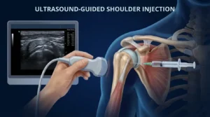
The Gold Standard: How Ultrasound Guidance Changes Everything
High-resolution musculoskeletal ultrasound functions like a real-time, high-definition window into your moving joint. It uses harmless, high-frequency sound waves to generate detailed images of your tendons, ligaments, muscles, and joint spaces during the evaluation.
When utilizing ultrasound guidance, the entire nature of a PRP procedure changes from an educated guess to a highly targeted delivery system.
1. Real-Time Visual Tracking
With the ultrasound probe placed gently on your shoulder, we can visually track the needle tip as it enters the skin and advances through the specific tissue layers. Every single millimeter of progress is visible on a high-definition monitor in real time.
2. Pinpoint Delivery
We can position the needle tip exactly inside the microscopic margins of a tendon tear, directly into the sheath of a painful biceps tendon, or into the precise layer of the glenohumeral joint space.
3. Avoidance of Vital Structures
Ultrasound images reveal blood vessels and nerves clearly. This allows the provider to chart a customized needle path that completely avoids delicate local structures, significantly reducing procedural discomfort and eliminating the risk of accidental nerve or vessel irritation.
4. Confirmation of Saturation
As the PRP is deployed, the fluid appears on the ultrasound monitor as a changing visual field. We can actually watch the plasma fill the tear or coat the targeted tissue, ensuring a complete therapeutic dose is left exactly where it can create the most impact.
According to a comprehensive clinical consensus from the American Academy of Orthopaedic Surgeons (AAOS), the use of image guidance significantly increases the baseline accuracy of joint and tendon injections across the body, directly correlating with improved patient outcomes and decreased procedural pain.
Maximizing Your Recovery After a PRP Injection
Because PRP triggers an intentional, localized inflammatory response to kickstart your body’s natural healing mechanisms, managing your recovery correctly is essential to securing an optimal long-term result. The recovery timeline for a shoulder PRP injection generally progresses through clear biological phases:
| Recovery Phase | Timeline | Primary Focus & Guidelines |
|---|---|---|
| Inflammatory Phase | Days 1–5 | Expect increased soreness or stiffness. This is a positive sign that platelets are actively releasing growth factors. Strictly avoid anti-inflammatory medications (NSAIDs) like ibuprofen or naproxen, as they can shut down the healing cascade. |
| Proliferative Phase | Weeks 2–4 | Initial procedural soreness subsides. The body begins laying down new, immature collagen fibers. Gentle, guided range-of-motion movements are introduced to help these fibers structurally align. |
| Remodeling Phase | Weeks 4–12+ | The new tissue matures, strengthens, and becomes increasingly resilient. Structured, customized physical therapy becomes vital to rebuild strength and stability for a safe return to sports. |
The Importance of a Dedicated Shoulder Evaluation
The shoulder is an exceptionally specialized joint that demands a matched level of clinical expertise. While many general wellness clinics, medspas, and broad pain management practices offer regenerative therapies today, a successful outcome always begins with an accurate mechanical diagnosis.
A structural tendon tear requires a completely different management strategy than advanced osteoarthritis, a labral tear, or a frozen shoulder. Imaging alone does not always provide the full picture; a high-resolution MRI or ultrasound must be paired with an expert physical exam that understands the nuances of overhead athletic movement.
An orthopedic shoulder specialist possesses an intimate, deep understanding of shoulder mechanics, advanced surgical alternatives, and specialized non-surgical treatments. They can comprehensively evaluate your condition to ensure that PRP is truly the right choice for your specific injury, and then execute the procedure with the strict spatial precision required for biological healing.
If you are dealing with persistent shoulder pain in the San Diego area and want to explore advanced, image-guided treatment options tailored to your active lifestyle, you can learn more by reviewing our comprehensive guide to Platelet-Rich Plasma (PRP) Injections in San Diego, CA.

This article is intended for general educational and informational purposes only and should not be considered medical advice. Reading this content does not create a physician-patient relationship. Always consult a qualified healthcare professional regarding your specific medical concerns, diagnosis, or treatment options.